Diagnostic Imaging & Radiology
Ashli Lafosse, Diagnostic Imaging Assistant Director; Kristi Hebert, RT, R, Diagnostic Imaging Director; Joan Wojak, M.D. Radiology Medical Director
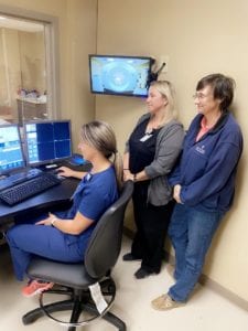
The Diagnostic Imaging Department operates 24 hours a day, 7 days per week, and provides a wide variety of diagnostic and therapeutic studies/procedures on patients of all ages (pediatric, infant, adolescent, adult, and geriatric) under the direction of Radiologists. All procedures are performed according to the guidelines set forth by the American College of Radiology and the standards of practice set forth by the department medical advisor.
The Diagnostic Imaging Department is located on the first floor adjacent to the Emergency Room, and is staffed with Board Certified Radiologists, Registered Licensed Radiologic Technologists, Registered Licensed CT Technologists, Registered Licensed Nuclear Medicine Technologists, Registered Licensed Mammography Technologists, Certified Ultrasonographers by ARDMS, and Clerical personnel.
General Radiography to include Fluoroscopy
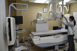 New General Electric Precision 500D Digital Radiographic & Fluoroscopic System:
The Fluoroscopic system is used for Upper & Lower GI exams, Myelography and Arthrography. This modality provides the Radiologist the ability to view the body part of interest in real time and depending on the exam, the body part in motion assisting in diagnoses. The Radiographic digital system is used to take X-rays of the human body in one dimension. With digital imaging the X-rays are emitted through the anatomical area of interest to the digital receptor, processing of the images is accomplished using computer software and specific algorithms for each body part. Viewing of the processed image is within 60 seconds.
Computerized Tomography
Abbeville General is pleased to announce the addition of a second CT scanner in the Diagnostic Imaging Department. The GE Revolution Evo is a 32-slice helical system with the following features: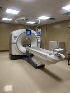
- 32 Channel / 64 Slice CT Scanner (can be referred to as a 64-slice scanner)
- Best-in-class Image quality (0.28mm spatial resolution) – helpful in seeing/diagnosing small lesions
- ASIR Low Dose Algorithm to reduce patient radiation dose by up to 50%
- Low-Dose CT Lung Screening protocol and indication for use
- Calcium Scoring to determine the amount of calcium in the coronary arteries, an important screening exam.
- Smart Metal Artifact Reduction – algorithm to remove metal caused from hip/knee implants/spine hardware, surgical clips for easier interpretation when metal is present
- High-Pitch Helical scan mode for imaging pediatrics and trauma patients
- Advanced in-room workflow to assist technologist during trauma imaging
- University of Wisconsin Dose Optimized Protocols to deliver the correct dose based on patient size and exam to be performed
- Ability to measure a stenosis in an artery and ability to remove bone to see vasculature better
Abbeville General Diagnostic Imaging will be able to offer a continuous provision of CT services for scheduled exams accommodating more patients, emergent exams, and eliminating delays especially during a biopsy procedure or a system failure/breakdown.
CT Cardiac Calcium Scoring
Abbeville General is now performing CT Cardiac Calcium Scoring. This heart scan is a non-invasive CT that provides pictures of your heart that can help detect and measure calcium-containing plaque in the arteries.
From this information, your doctor can identify possible coronary artery disease before you have signs and symptoms.
Your doctor will then use your test results to determine if you may need medication or lifestyle changes to reduce the risk of heart disease.
To schedule an appointment, you will need to contact your physician to obtain an order for your CT Cardiac Calcium Scoring. Once you have the physician’s order, call Centralized Scheduling at 898-6519 to schedule your appointment.
For more information on CT Cardiac Calcium Scoring, CLICK HERE.
To watch a video on CT Cardiac Calcium Scoring, CLICK HERE.
Computed Tomography (CT) – Chest
Computed tomography (CT) of the chest uses special x-ray equipment to examine abnormalities found in other imaging tests and to help diagnose the cause of unexplained cough, shortness of breath, chest pain, fever and other chest symptoms. CT scanning is fast, painless, noninvasive and accurate. Because it is able to detect very small nodules in the lung, chest CT is especially effective for diagnosing lung cancer at its earliest, most curable stage.
Tell your doctor if there is a possibility you are pregnant and discuss any recent illnesses, medical conditions, medications you are taking, and allergies.
You will be instructed not to eat or drink anything for a few hours beforehand.
If you have a known allergy to contrast material, your doctor may prescribe medications to reduce the risk of an allergic reaction. These medications must be taken beginning 12 hours prior to your exam.
Leave jewelry at home and wear loose, comfortable clothing. You may be asked to wear a gown.
If your Physician orders IV Contrast, an IV will need to be started.
Blood work may be done prior to IV contrast administration to check how well your kidneys are working.
For more information, CLICK HERE.
Head – Computed Tomography (CT)
Computed tomography (CT) of the head uses special x-ray equipment to help assess head injuries, severe headaches, dizziness, and other symptoms of aneurysm, bleeding, stroke, and brain tumors. It also helps your doctor to evaluate your face, sinuses, and skull or to plan radiation therapy for brain cancer. In emergency cases, it can reveal internal injuries and bleeding quickly enough to help save lives.
Tell your doctor if there’s a possibility you are pregnant and discuss any recent illnesses, medical conditions, medications you’re taking, and allergies.
You will be instructed not to eat or drink anything for a few hours beforehand. If you have a known allergy to contrast material, your doctor may prescribe medications to reduce the risk of an allergic reaction.
Leave jewelry at home and wear loose, comfortable clothing. You may be asked to wear a gown.
If your Physician orders IV Contrast, an IV will need to be started.
Blood work may be done prior to IV contrast administration to check how well your kidneys are working.
For more information, CLICK HERE.
Abdomen and Pelvis- Computed Tomography (CT)
Computed tomography (CT) of the abdomen and pelvis is a diagnostic imaging test used to help detect diseases of the small bowel, colon and other internal organs and is often used to determine the cause of unexplained pain. CT scanning is fast, painless, noninvasive and accurate. In emergency cases, it can reveal internal injuries and bleeding quickly enough to help save lives.
Tell your doctor if there’s a possibility you are pregnant and discuss any recent illnesses, medical conditions, medications you’re taking, and allergies.
If you have a known allergy to contrast material, your doctor may prescribe medications to reduce the risk of an allergic reaction. These medications must be taken 12 hours prior to your exam. Leave jewelry at home and wear loose, comfortable clothing. You may be asked to wear a gown.
You will be instructed not to eat or drink anything for a few hours beforehand.
If your Physician orders IV Contrast, an IV will need to be started.
CT Abdomen and Pelvis: If Oral contrast is ordered by your Physician for you will need to consume Two 16 ounce cups of diluted flavored Iodinated oral contrast. CT will be done 90 minutes after you have completed consuming oral contrast.
CT Abdomen only: If Oral contrast is ordered by your Physician, you will need to consume One 16 ounce cup of diluted flavored Iodinated oral contrast. CT will be done 30 minutes after you have completed consuming oral contrast.
Blood work may be done prior to IV contrast administration to check how well your kidneys are working.
For more information, CLICK HERE.
Nuclear Medicine
![]()
Nuclear Medicine
Nuclear medicine imaging uses small amounts of radioactive materials called radiotracers that are typically injected into the bloodstream, inhaled or swallowed. The radiotracer travels through the area being examined and gives off energy in the form of gamma rays which are detected by a special camera and a computer to create images of the inside of your body. Nuclear medicine imaging provides unique information that often cannot be obtained using other imaging procedures and offers the potential to identify disease in its earliest stages.
Tell your doctor if there’s a possibility you are pregnant or if you are breastfeeding and discuss any recent illnesses, medical conditions, allergies and medications you’re taking. Depending on the type of exam, your doctor will instruct you on what you may eat or drink beforehand, especially if sedation (anesthesia) is to be used. Leave jewelry at home and wear loose, comfortable clothing. You may be asked to wear a gown.
For more information, CLICK HERE.
Planar Imaging
- Whole Body Bone Scan
- Ventilation & Perfusion Lung Scan
- HIDA Scan – Liver and Gallbladder Imaging
- Gastric Emptying
- Liver and Spleen Imaging
- Tumor Imaging
- Thyroid Uptakes
- MUGA Scan
- SPECT Cardiac Perfusion Studies (MPI) – Exercise Stress and Medication Stress (Lexiscan). Click here for more information.
Hepatobiliary Scan (HIDA) – Nuclear Medicine
Hepatobiliary nuclear medicine imaging helps evaluate the parts of the biliary system, including the liver, gallbladder and bile ducts, using small amounts of radioactive materials called radiotracers that are typically injected into the bloodstream.
Do not eat or drink anything at least 4 hours prior to your scan. You may take your scheduled medication with a sip of water only.
Tell your doctor if there’s a possibility you are pregnant or if you are breastfeeding and discuss any recent illnesses, medical conditions, allergies and medications you’re taking. Leave jewelry at home and wear loose, comfortable clothing. You may be asked to wear a gown.
For more information, CLICK HERE.
Skeletal Scintigraphy (Bone Scan)
Skeletal scintigraphy helps to diagnose and evaluate a variety of bone diseases and conditions using small amounts of radioactive materials called radiotracers that are injected into the bloodstream. The radiotracer travels through the area being examined and gives off radiation in the form of gamma rays which are detected by a special gamma camera and a computer to create images of your bones. Because it is able to pinpoint molecular activity within the body, skeletal scintigraphy offers the potential to identify disease in its earliest stages.
Tell your doctor if there’s a possibility you are pregnant or if you are breastfeeding. Discuss whether you have recently taken a bismuth-containing medicine such as Pepto-Bismol or had a barium contrast x-ray. Your doctor will instruct you on how to prepare and will likely tell you to drink extra fluids after the radiotracer is injected. Leave jewelry at home and wear loose, comfortable clothing. You may be asked to wear a gown.
You will be questioned concerning recent illnesses, medical conditions, allergies and home medication, including vitamins and herbal supplements.
An IV will be started and the radioactive tracer will be administered.
You will be instructed to return in 4 hours for the Scan.
The scan time will be approximately 20 minutes when you return.
For More Information, CLICK HERE.
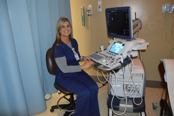
Ultrasound
 Abdominal Imaging, including, but not limited to:
- Gallbladder
- Liver
- Spleen
- Kidney
- OB/GYN
- Breast
- Thyroid
Vascular:
- Arterial / Venous Doppler Imaging
Pelvis Ultrasound
Ultrasound imaging of the pelvis uses sound waves to produce pictures of the structures and organs in the lower abdomen and pelvis. There are three types of pelvic ultrasound: abdominal, vaginal (for women), and rectal (for men). These exams are frequently used to evaluate the reproductive and urinary systems. Ultrasound is safe, noninvasive and does not use ionizing radiation.
One hour prior to Ultrasound procedure you will need to drink 32-48 ounces of water to assure a full urinary bladder. A full urinary bladder helps to lift the intestines, allowing for easier visualization of your pelvic organs.
This procedure requires little to no special preparation. Leave jewelry at home and wear loose, comfortable clothing. You may be asked to wear a gown.
For more information, CLICK HERE.
Abdomen – Ultrasound
Ultrasound imaging of the abdomen uses sound waves to produce pictures of the structures within the upper abdomen. It is used to help diagnose pain or distention (enlargement) and evaluate the KIDNEYS, LIVER, GALLBLADDER, bile ducts, PANCREAS, SPLEEN and ABDOMINAL AORTA. Ultrasound is safe, noninvasive and does not use ionizing radiation.
Do not eat or drink anything at least 6 hours prior to your Ultrasound. You may take your scheduled medication with a sip of water only.
Leave jewelry at home and wear loose, comfortable clothing. You may be asked to wear a gown.
For more information, CLICK HERE.
Breast – Ultrasound
Ultrasound imaging of the breast uses sound waves to produce pictures of the internal structures of the breast. It is primarily used to help diagnose breast lumps or other abnormalities your doctor may have found during a physical exam, mammogram or breast MRI. Ultrasound is safe, noninvasive and does not use radiation.
This procedure requires little to no special preparation. Leave jewelry at home and wear loose, comfortable clothing. You will be asked to undress from the waist up and to wear a gown during the procedure.
For more information, CLICK HERE.
Venous (Extremities) – Ultrasound
Venous ultrasound uses sound waves to produce images of the veins in the body. It is commonly used to search for blood clots, especially in the veins of the leg – a condition often referred to as deep vein thrombosis. Ultrasound does not use ionizing radiation and has no known harmful effects.
On occasion, you may be asked not to eat or drink anything but water for six to eight hours beforehand. Otherwise, little or no special preparation is required for this procedure.
Leave jewelry at home and wear loose, comfortable clothing. You may be asked to wear a gown.
For more information, CLICK HERE.
Magnetic Resonance Imaging (MRI)
Magnetic Resonance Imaging (MRI)
Magnetic resonance imaging (MRI) uses a powerful magnetic field, radio waves and a computer to produce detailed pictures of the body’s internal structures that are clearer, more detailed and more likely in some instances to identify and accurately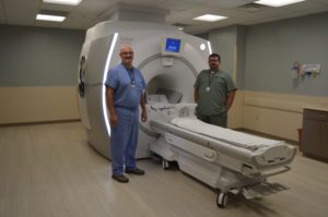 characterize disease than other imaging methods. It is used to evaluate the body for a variety of conditions, including tumors and diseases of the liver, heart, and bowel. MRI is noninvasive and does not use ionizing radiation.
Our NEW GE 3 Tesla MRI offers whole-body imaging including Magnetic Resonance Angiography (MRA). This unit is accredited by the American College of Radiology.
For information on Magnetic Resonance Imaging (MRI) Safety, CLICK HERE.
Trophon EFR
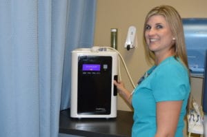 The Trophon is an environmentally safe system providing high level disinfection of the probe shaft and handle providing consistent quality assurance. The in-process chemical indicator and a unique quality sensor validates each disinfection cycle is completed. The disinfection cycle takes place within seven minutes in a fully automated closed system preventing staff and patients from exposure to toxic, harmful chemicals. Small quantities of water and oxygen are generated as by products. There are three units placed in the ultrasound exam rooms that perform OB procedures.
Breast Biopsies and Drainage Procedures
Breast Biopsy, Ultrasound-Guided Fine Needle Aspiration
An ultrasound-guided breast biopsy uses sound waves to help locate a lump or abnormality and remove a tissue sample for examination under a microscope. It is less invasive than surgical biopsy, leaves little to no scarring and does not involve exposure to ionizing radiation.
Day of procedure, upon arrival in Diagnostic Imaging Department, blood will be collected to determine how well your blood is clotting and an assessment by a Nurse will be performed. It typically takes an hour for blood test results to be completed.
Tell your doctor about any recent illnesses or medical conditions and whether you have any allergies, especially to anesthesia. Discuss any medications you’re taking, including herbal supplements and aspirin. You will be advised to stop taking aspirin or blood thinner three days before your procedure. Leave jewelry at home and wear loose, comfortable clothing. You may be asked to wear a gown. If you are to be sedated, plan to have someone drive you home afterward.
For more information, CLICK HERE.
Thyroid Biopsy, Ultrasound-Guided Fine Needle Aspiration
An ultrasound-guided fine needle aspiration biopsy uses sound waves to help locate a nodule or abnormality within the thyroid and remove a tissue sample for examination under a microscope. The procedure is less invasive than surgical biopsy, leaves little to no scarring and does not involve exposure to ionizing radiation.
Day of procedure upon arrival in Diagnostic Imaging Department, blood will be collected to determine how well your blood is clotting and an assessment by a Nurse will be performed. It typically takes an hour for blood test results to be completed.
This procedure requires little to no special preparation. Tell your doctor about any medications you’re taking, such as aspirin or blood thinners.
For more information, CLICK HERE.
Thoracentesis
Thoracentesis uses imaging guidance and a needle to help diagnose and treat pleural effusions, a condition in which the space between the lungs and the inside of the chest wall contains excess fluid. It is performed to help determine the cause of the excess fluid and to ease any shortness of breath or pain by removing the fluid and relieving pressure on the lungs.
Day of procedure upon arrival in Diagnostic Imaging Department, blood will be collected to determine how well your blood is clotting and an assessment by a Nurse will be performed. It typically takes an hour for blood test results to be completed.
Your doctor will instruct you on how to prepare, including any changes to your medication schedule. Tell your doctor if there’s a possibility you are pregnant and discuss any recent illnesses, medical conditions, allergies and medications you’re taking, including herbal supplements and aspirin. You may be advised to stop taking aspirin, nonsteroidal anti-inflammatory drugs (NSAIDs), or blood thinners several days prior to your procedure. Leave jewelry at home and wear loose, comfortable clothing. You may be asked to wear a gown.
For more information, CLICK HERE.
Radiologist Team
Our focus is YOU, and our goal is to deliver excellence in diagnostic imaging care.
Acadiana Radiology Group is a Lafayette based group practice that provides professional radiology services to nine healthcare facilities in the Acadiana area. Its members maintain specialized training and experience in adult and pediatric diagnostic radiology, interventional radiology, neuroradiology and mammography.
Radiologists

Dr. Doug Acomb
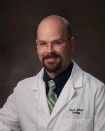
Dr. Paul Billeaud

Dr. Patrick Boyle

Dr. Kyle Degeyter
Dr. Blaine Hoppe

Dr. Mark T. Stephan
Dr. Joan Wojak
Medical Director of Radiology
at Abbeville General